9 easy steps for an oral cancer examination
According to studies that have been conducted over the years, 45,000 cases of squamous cell carcinoma in the oral cavity and the throat are reported in the United States every year. As a result, 8,000 deaths from the condition are reported each year. Diagnosis of the condition in its early stages plays a major role in the success rate of its treatment. Despite advanced treatment opportunities available in the field now, the 5-year survival rate of patients in advanced or moderate stages is less than 60%. Therefore, it is immensely important to diagnose oral cancer in its early stages, for which a thorough oral cancer examination is needed. A screening at every dental visit can catch all problems at their nascent stage and help you get timely treatment.
The oral cancer examination procedure can be divided into two parts—the extraoral examination and the intraoral examination. Here’s the complete procedure explained in 9 steps.
Extra-oral examination
- Face
Any asymmetries, mass development, swelling, or discoloration on the face is examined. Any pigmented, ulcerated, raised, or firm areas of the skin of the face and scalp is also screened. - Eyes
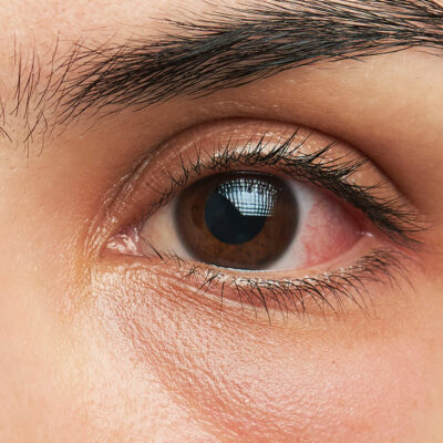
The extraocular movement of the eye is checked in every direction in order to examine the cranial nerve. The eyes and the periorbital area are to be checked for any swelling, which can be a sign of a tumor. - Ears and nose
During the nasal examination, the external nose and the paranasal area overlying the maxilla and maxillary sinus are examined. To check the acoustic nerve in the ear, normal talking is done with the patient. - Neck
The neck is manually palpated to check and compare both sides of the neck for any signs of enlargement of lymph nodes. - Thyroid
The thyroid gland is first examined and then palpated because it is difficult to feel. The patient can be asked to swallow while the doctor has his fingers placed adjacent to the thyroid gland to make the examination thorough. Swallowing will make any kind of abnormality visible to the examiner. - Lips
The lips are examined with mouth open and mouth closed. Any abnormalities in the color, symmetry, contour, or texture of the lips can raise an alarm.
Intraoral examination
- Buccal mucosa
The buccal mucosa is moved away from the teeth and gingiva to check the vestibule, one side at a time. Although the appearance of a white line is normal, any other abnormalities with the texture or color can point towards an issue. - Tongue
The patient is asked to put their tongue out and move it in both the directions. The movement should be smooth and there should be no spasm or asymmetry. Presence of any masses, ulceration, or swelling is noted. - Hard and soft palate
The patient is asked to keep their mouth wide open and their head tilted backward. Presence of loose teeth, white spots, red spots, ulcerations, rough areas, asymmetry, or any masses can be a sign of cancer in the mouth, head, or neck.


